Michigan Retina-Vitreous Institute
1290 South Linden Road, Flint, Michigan 48532
(810) 732-6231
Examination
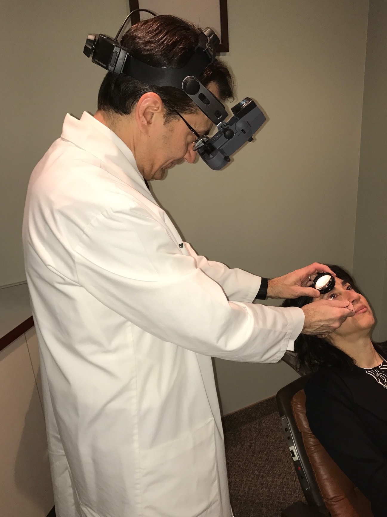
Early detection and treatment holds the best promise for treating many retinal disorders. This involves detailed examination, often times the use of sophisticated testing services, detailed explanations of the findings and treatment options, and advanced medical and surgical treatments.
Examination - Thorough examination of vitreous and retinal disorders can involve numerous detailed steps. Detailed medical histories are taken, measurements of visual function are taken, and examination of the front of the eyes, scleral depressed examination of the back of the eye, contact lens examination of the back of the eye, and directed general physical examination can all be involved.
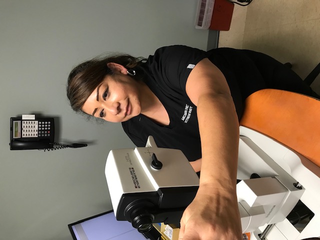
Testing
Fundus Photography - Specialized digital cameras are used to document abnormalities in the retina. This is key in following the progress of certain retinal disease and to monitor treatment.
Fluorescein and Indocyanine Green Angiography - An intravenous injection of diagnostic dye is given in the arm or hand, after which numerous high definition digital photographs are taken of the retina. This commonly performed testing provides some of the most useful anatomic and functional information of the retina which can be of paramount importance in the diagnosis and treatment of retinal disorders.
Optical Coherence Tomography - Using infrared light computer controlled scanning of the eye, very fine cross-sectional detailed images of the macula can be obtained which aid in diagnosis and treatment decisions.
Ocular Ultrasonography - Through the use of high frequency sound waves, the structures of the back of the eye can be visualized and evaluated when direct visualization is impaired, such as through cataract or blood in the eye. In addition to this Diagnostic B-scan, Diagnostic A-scan ultrasonography can evaluate the internal characteristics of tumors and other lesions in the eye.
Visual Field Testing (perimetry) - Computer controlled perimetry maps the peripheral or side vision, which provides useful information in diagnosing and monitoring certain disorders of the eye.
Visual Electrophysiology - Detailed information on retinal function is obtained through the use of computer assisted amplification and analysis of the electrical waves produced by the eyes. Full field electroretinography (ERG) tests the function of the rods and cones in the retina in such conditions as retinitis pigmentosa, other retinal dystrophies, and certain circulatory disorders of the retina. Electrooculography (EOG) measures the function of the retinal pigment epithelium and is useful in classifying certain dystrophies and other disorders of the retina.
Treatment
Medications - Certain diseases of the retina and vitreous can be treated with medications, either in eye drop form, pills, or injections into the eye.Laser Surgery -Laser retinal surgery is sometimes needed to treat conditions such as diabetic retinopathy, age-related macular degeneration, retinal tears, retinal vein occlusions, and other conditions. Such treatments are performed in the office.
Surgery - Operating room surgical procedures are done on an outpatient basis at local facilities. Scleral buckling surgery for the repair of retinal detachment and posterior vitrectomy surgery for advanced diabetic retinopathy, retinal detachment, macular holes, epiretinal macular membranes, and a host of other conditions are commonly performed. Certain procedures, such as injections of medications for macular degeneration or edema and pneumatic retinopexy for repair of retinal detachment, can be performed in the office.
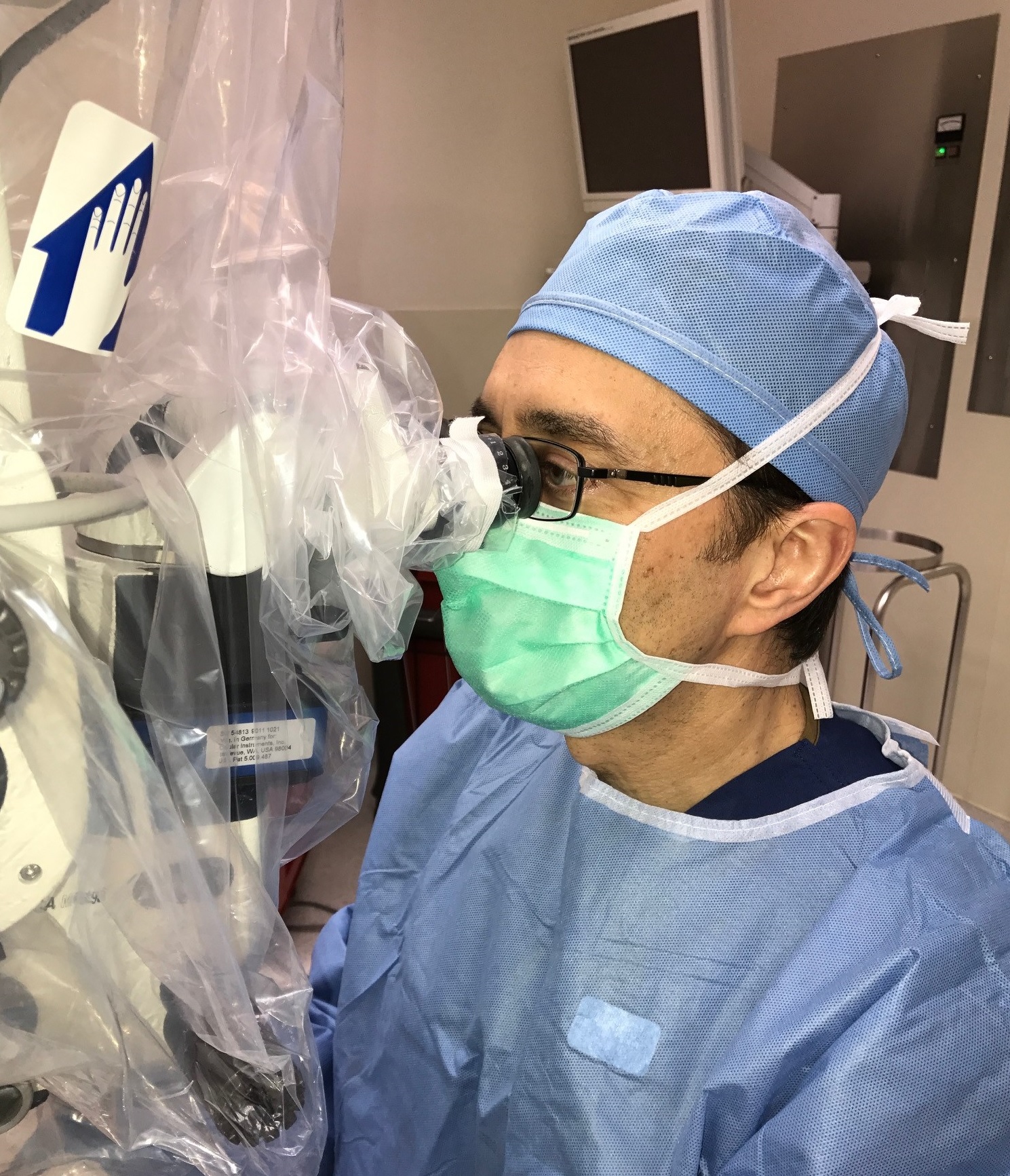
Most surgeries are performed at The Surgery Center, the area's first, most experienced, and only physician-controlled multi-specialty ambulatory surgery center. At times, surgeries are performed at local area hospitals.
VITRECTOMY SURGERY
Vitrectomy surgery is a microsurgical technique that is used to treat a variety of retinal diseases. In this operation, three small needle-sized incisions are made through the eye wall allowing the removal of the vitreous gel that fills the majority of the eye. This allows for clearing of blood, the removal of scar tissue, and the repair of detachment. Laser treatment may be applied, and in some cases, a gas or silicone oil bubble may be left in the eye to hold your retina in place while it heals. Should a gas bubble be used, you cannot fly in an airplane or undergo anesthesia utilizing nitrous oxide gas until the bubble evaporates in 3 to 8 weeks. In addition, you may need to position your head appropriately (sometimes directly face down) to allow for proper healing. More than one operation are sometimes needed.
SCLERAL BUCKLE
This surgery involves the placement of a silicone band around the equator of the exterior of the eye underneath the muscles that control the movement of the eye. This indents the eye wall compressing tears and flattening a retinal detachment. Sometimes a small gas bubble is injected into the eye and sometimes this technique is combined with vitrectomy surgery to repair more complicated retinal detachments.